
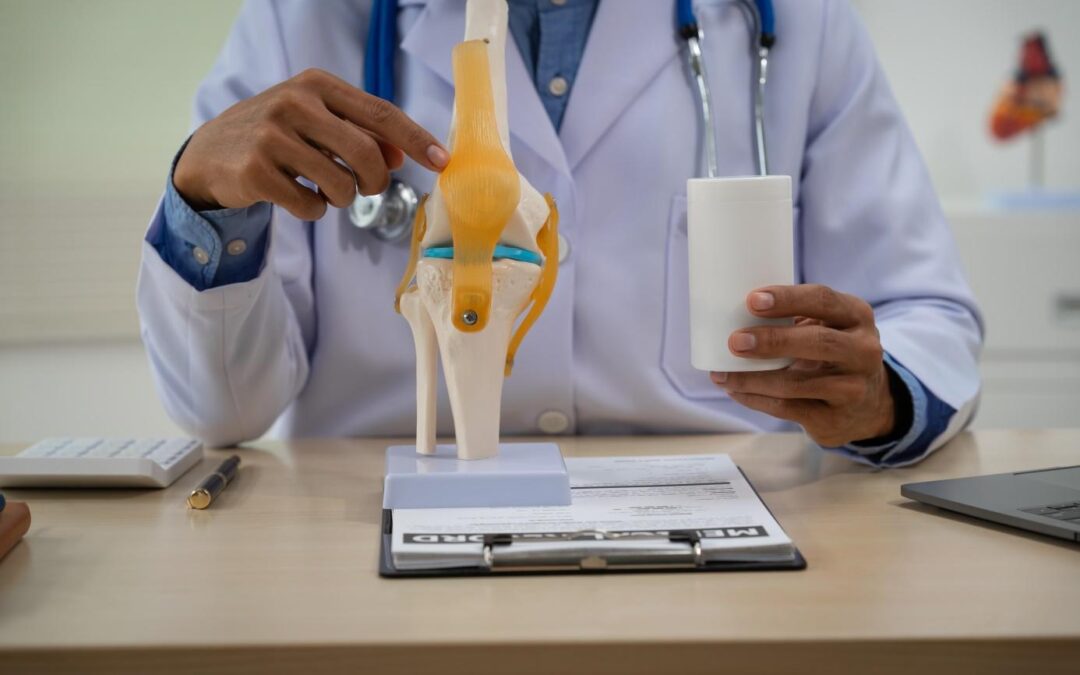
Medial Collateral Ligament: Understanding, Preventing, and Relieving
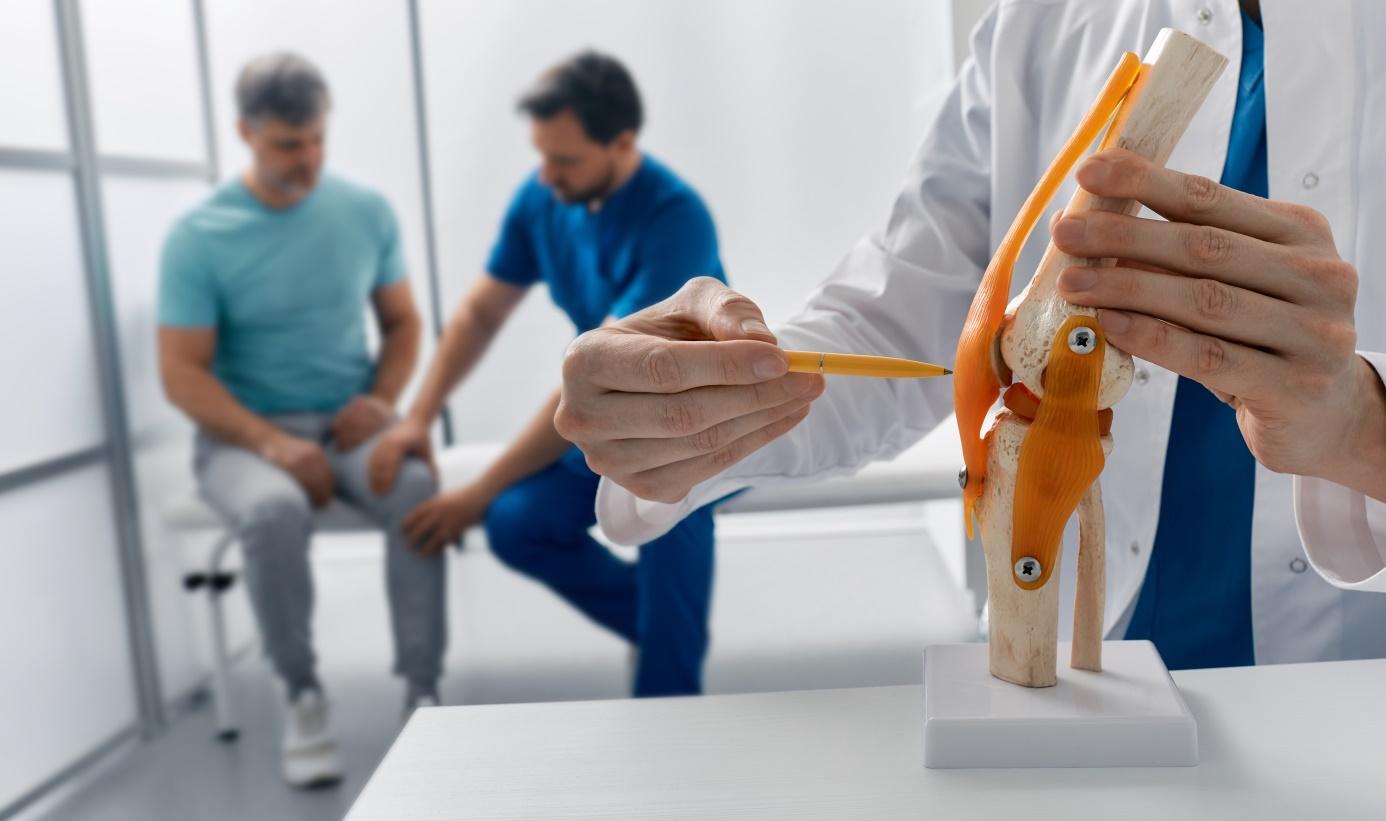
The knee is a complex and vital joint, essential for our daily mobility, whether for walking, running, or engaging in sports activities. At the core of its stability lies a network of ligaments, among which the medial collateral ligament, or MCL, also known as the internal collateral ligament (ICL), stands out. Its primary function is to stabilize the knee and prevent excessive inward-to-outward movements, thereby ensuring the joint’s cohesion. Injuries to the medial collateral ligament are the most common ligament injuries of the knee. Physiotherapy plays a crucial role in the healing and rehabilitation of these injuries, allowing patients to regain their normal function.
What is a Medial Collateral Ligament Tear?
An MCL tear occurs when this ligament is excessively stretched or torn. It can result from a direct blow to the knee, a sudden change in direction, or a “forced valgus” trauma. The latter mechanism is common: the foot is planted on the ground, and the knee experiences pressure from the outside toward the inside, placing intense tension on the internal ligament. The ligament may either resist or rupture. The tear can be partial or complete, affecting one or more bundles of ligament fibers. Injuries are often more severe if the trauma occurs on a straight leg.
MCL Tears are Classified into Three Grades of Severity:
- Mild: involves slight stretching or a minimal tear of the ligament,
- Moderate: corresponds to a partial tear of the MCL,
- Severe: a complete tear of the ligament.
Despite the apparent seriousness of complete tears, it is essential to know that the medial collateral ligament has a good healing potential, even in the case of total rupture.
What Are the Symptoms of an MCL Tear?
Recognizing the symptoms of an MCL tear quickly is essential for early diagnosis and treatment. Common signs include:
- Pain: generally located on the inner side of the knee, i.e., the side toward the other knee. The pain can be sharp and occur immediately after the injury.
- Swelling and inflammation: Swelling may appear shortly after the injury, indicating tissue inflammation. Swelling can be rapid or delayed depending on associated injuries.
- Bruising or contusion: Bruises may develop within hours or days after the injury. They are often visible on the inner side of the knee and may extend down the leg. The extent of bruising depends on the severity of the injury.
- A feeling of instability: the patient may experience instability or a sensation that the knee feels like it is going to give way, particularly during weight-bearing activities on the affected leg. A sensation of the knee “dislocating” inward may also be present.
- Limited range of motion: The injury often results in stiffness and a reduced ability to extend or flex the knee fully.
- A popping sound: it may be heard or felt at the moment of the accident.
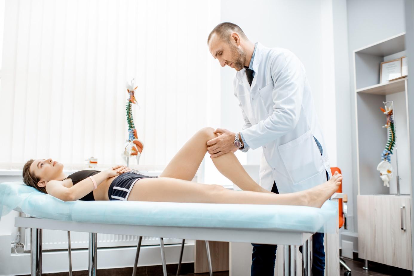
How is a Rupture Diagnosed?
An accurate diagnosis is crucial for determining the most suitable treatment plan. The diagnostic process generally involves:
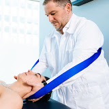
- Clinical examination: A physiotherapist will perform a thorough examination of the knee. They will check for tenderness along the MCL, swelling, and instability. The physiotherapist may perform specific tests, such as stabilizing the knee and gently pulling the leg outward to check for abnormal movement or an “internal opening” of the joint.
- If the opening appears only when the knee is bent, it suggests a partial rupture.
- If laxity exists in both flexion and extension, it indicates a complete rupture.
- The position of the knee and the body’s movement at the time of injury can also aid in diagnosis.
- Imaging studies:
- X-rays help rule out bone fractures.
- Magnetic Resonance Imaging (MRI) is essential for confirming the diagnosis, as it provides detailed images of soft tissues, including the MCL. MRI also helps detect associated injuries since MCL tears are rarely isolated and often occur alongside an anterior cruciate ligament (ACL) rupture.
How Long Can the Pain Last?
The recovery time after a medial collateral ligament (MCL) tear depends on the severity of the injury and the effectiveness of the treatment plan. Here are the general timeframes:
- Mild tear: recovery usually takes between 1 and 3 weeks. Patients are encouraged to gradually resume normal activities while avoiding excessive stress on the knee.
- Moderate tear: recovery generally lasts between 4 and 6 weeks. Physiotherapy plays a crucial role during this period in restoring full function and preventing recurrence.
- Severe tear: Healing can take from 8 to 12 weeks or longer. Intensive rehabilitation and, in some cases, surgery are necessary for optimal recovery.
Treatments
Treatment of an MCL tear varies depending on its severity. Many MCL tears can be managed non-surgically through conservative approaches.
How to Relieve the Tear?
Managing pain and inflammation is a priority. Treatments include:
- Medications: Non-steroidal anti-inflammatory drugs (NSAIDs) can be used to relieve pain. For persistent pain that impairs function, a local corticosteroid injection may be a suitable treatment option.
- Physiotherapy: A physiotherapist will design a specific exercise program for rehabilitation.
- Improving mobility and flexibility: Manual therapy techniques, stretching, and mobility exercises are used to restore normal knee range of motion and prevent joint stiffness.
- Muscle strengthening: targeted exercises on muscles around the knee are essential to stabilize the joint and prevent excessive movements that could worsen the injury.
- Functional rehabilitation: Once pain is reduced and strength, mobility, and flexibility are improved, the physiotherapist focuses on the gradual reintegration of the patient’s daily activities, such as sports or professional pursuits, while aiming to prevent relapse and promote full recovery.
- Manual therapy techniques: these can also be used to relieve muscle tension and spasms associated with the injury.
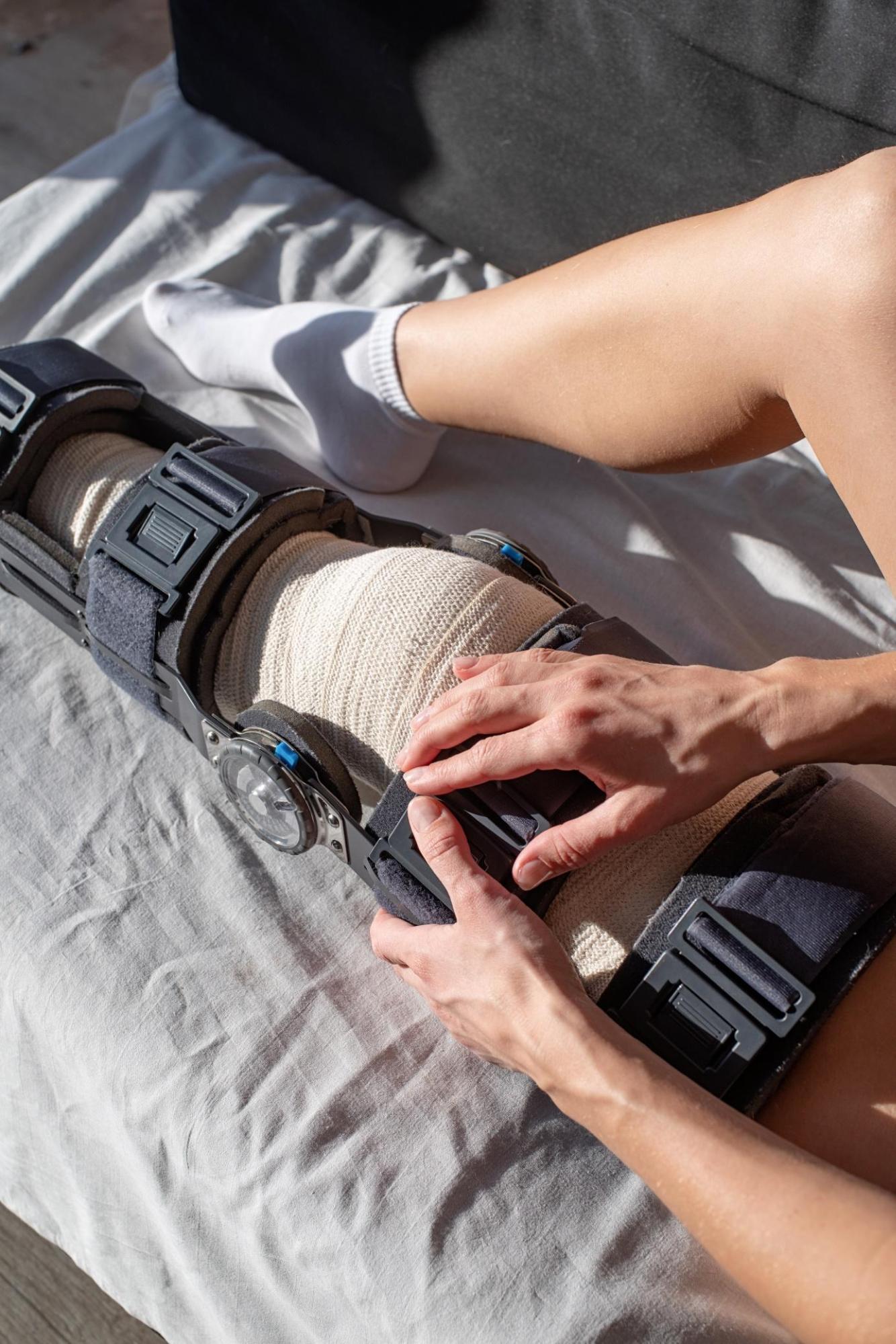
What Are the Benefits of a Brace?
The use of a brace or hinged knee support is often recommended in non-surgical treatment, particularly for moderate to severe tears, as well as after surgery. Its advantages include:
- Support and stabilization: The brace provides support to the knee, helping stabilize the joint.
- Prevention of further injury: it protects the ligament during its healing phase.
- Dynamic immobilization: unlike a non-hinged splint that fully immobilizes the leg, a hinged knee brace allows bending and straightening movements. This dynamic immobilization is crucial because it promotes the effective healing of ligament fibers while reducing the risk of joint stiffness.
- Facilitation of early rehabilitation: The brace enables functional treatment to commence as soon as possible after the sprain, which is crucial in preventing stiffness and promoting a faster recovery.
- Long-term protection: it should be worn for a defined period (e.g., three weeks day and night, then three weeks during the day, totaling six weeks). For athletes or those returning to high-risk activities, such as skiing, the use of a specific 4-point brace may be recommended for up to one year after surgery or resumption of sports.
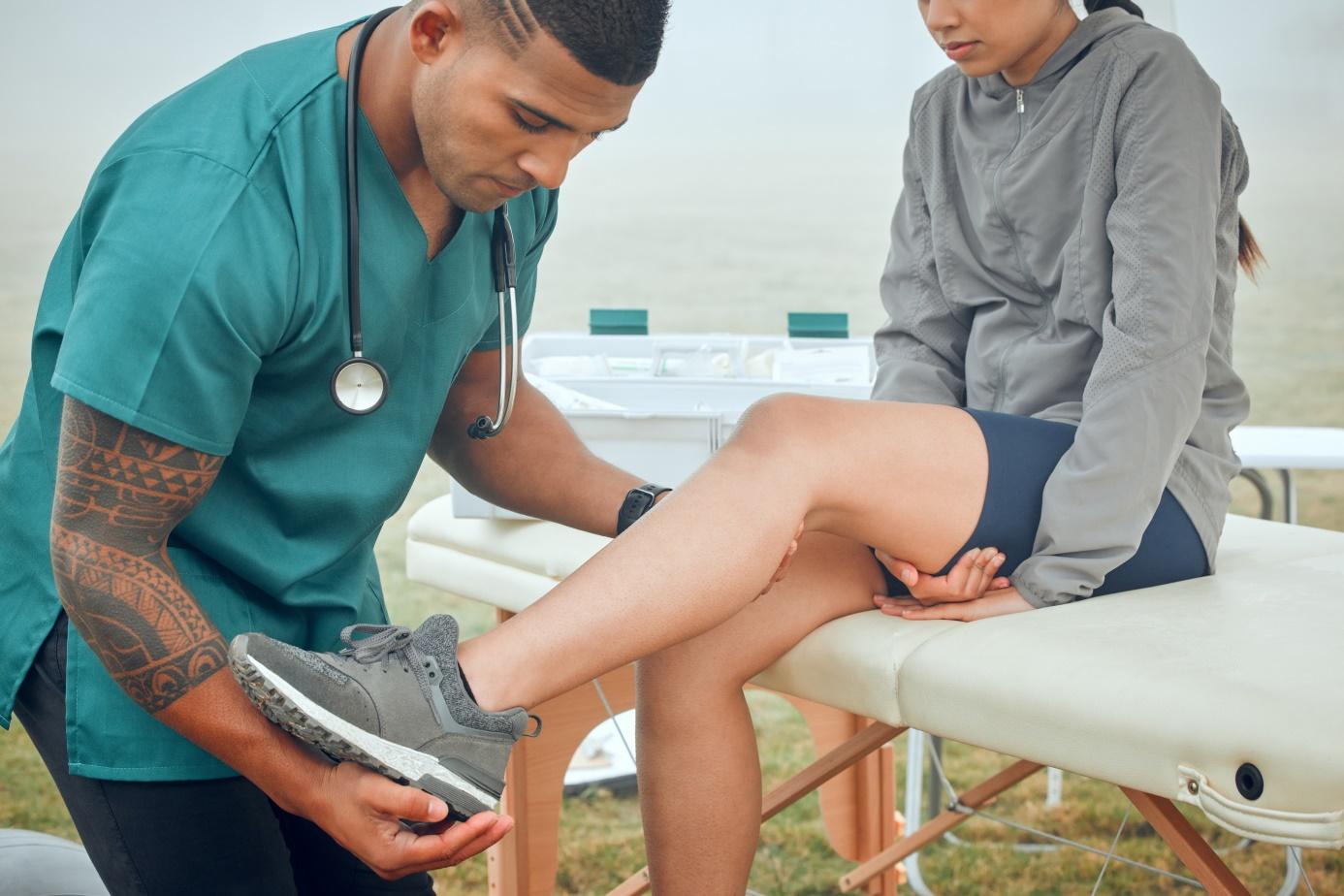
What Are the Benefits of Consulting a Specialist?
Seeing a qualified physiotherapist or sports doctor is essential as soon as the first signs of a knee injury appear. A healthcare professional can:
- Assess the extent of the injury by performing precise tests and evaluations to determine the degree of damage and affected structures, which is crucial for an accurate diagnosis.
- Develop a personalized treatment plan: Tailor the rehabilitation program to the patient’s specific needs and the severity of their injury.
- Guide rehabilitation: physiotherapists support the patient at every step of the healing process, from initial pain management to reintegration of activities.
- Prevent future injuries: Beyond treating the current injury, physiotherapy plays a key role in preventing recurrence. The physiotherapist can provide advice on safe movement techniques, injury prevention exercises, and activity modifications to reduce risks. Learning to land properly (e.g., on a varus knee rather than a valgus knee) is an essential aspect of prevention and rehabilitation.
- Determine the need for surgery: the surgeon will evaluate whether an operation is necessary, especially in complex or multi-ligament cases, and plan pre-operative rehabilitation.
Conclusion
A medial collateral ligament tear is a common knee injury that often responds very well to conservative treatment and physiotherapy rehabilitation. By combining effective pain management, targeted muscle strengthening, improved mobility, and progressive functional rehabilitation, physiotherapists can help patients regain normal function and resume daily and sports activities without pain or limitations. Prevention and consulting a physiotherapist are the keys to a successful and lasting recovery.
Author
-
We are physiotherapists passionate about movement and rehabilitation, with a clear goal: helping people better understand their pain and return to an active, unrestricted life.
Through our practice and content, we share practical, science-based approaches to prevent injuries, relieve pain, and sustainably improve mobility. We believe that well-informed patients make better decisions and achieve better outcomes.

Richard Bouzaglou, B.Sc. PT
Physiotherapist | Co-Founder
An experienced professional, Richard is the co-founder of the AMS Medical and Rehabilitation Center, where he has played a key role since 2008. With a background in sports medicine and physiotherapy, he has developed strong clinical expertise, particularly working with elite athletes.
His practice is based on a comprehensive approach that integrates advanced manual therapy, functional rehabilitation, and personalized care. Committed to continuous education and mentoring students, he is known for his clinical rigor and his ability to build lasting, trust-based relationships with his patients.

Moshe Vazana
Physiotherapist
With over 15 years of experience, Moshe is recognized for his precise, evidence-based approach. A physiotherapy graduate with advanced training in several methodologies, including the McKenzie Method (MDT) and the Mulligan Concept, he specializes in treating musculoskeletal and spinal conditions.
His international background and commitment to excellence enable him to provide care that is tailored, effective, and sustainable. Passionate about knowledge sharing, he is also actively involved in training the next generation of physiotherapy professionals.
Recent Comments