The shoulder joint is the most mobile in the human body, but also the most vulnerable to dislocations, accounting for nearly 50% of all major joint dislocations. Recognizing the signs of a glenohumeral dislocation and seeking urgent medical care are key to recovery and minimizing the risk of long-term damage.
What Is a Shoulder Dislocation and a Glenohumeral Dislocation?
A shoulder dislocation refers to the joint bones moving out of place. The most common type is the glenohumeral dislocation, often described as the “classic dislocated shoulder.”
Specifically, this happens when the head of the humerus (upper arm bone) slips out of the glenoid cavity, the cup-like socket of the scapula (shoulder blade). Think of it as a golf ball (humeral head) slipping off its tee (glenoid). This design allows wide mobility but also creates natural instability.
Dislocation can damage surrounding tissues, including muscles, nerves, tendons, ligaments, and blood vessels.
- Over 95% of cases are anterior dislocations (forward).
- Less common are posterior or inferior dislocations (the latter called luxatio erecta).
Causes of Glenohumeral Dislocation
Most shoulder dislocations are traumatic, resulting from sudden force. Common causes include:
- Hard falls on an outstretched arm or directly on the shoulder.
- Motor vehicle accidents.
- Contact sports or abrupt arm movements.
- Anterior dislocations: often caused by forced abduction with external rotation.
- Posterior dislocations: may result from seizures, electrical shock, or electrotherapy without muscle relaxants.

Symptoms of a Glenohumeral Dislocation
Five main warning signs:
- Severe, sudden pain: sharp and throbbing, worsens with any attempt to move. Muscle spasms often develop, further restricting movement.
- Visible deformity: the shoulder may look flattened or squared, instead of round. The arm may hang lower, appear longer, or sit at an unusual angle. Swelling and bruising develop quickly.
- Restricted movement: the arm cannot be moved without intense pain; daily tasks like dressing or grooming become impossible.
- Numbness and tingling (paresthesia): nerve compression (axillary nerve, brachial plexus) may cause tingling in the arm, hand, or fingers. This is a red flag for possible nerve injury.
- Swelling and bruising: inflammation appears within minutes, while bruising changes colour over days (red/purple → green/yellow).
How Is It Diagnosed?
Diagnosis starts with clinical presentation and injury history, confirmed with imaging:
- X-rays: multiple views (AP, axillary, Y-view) confirm dislocation, assess humeral head displacement, and check for fractures (humeral neck, glenoid, greater tuberosity).
- MRI: especially for young patients (labrum integrity) and older patients (rotator cuff tears).
- CT scans: useful for recurrent dislocations, evaluating bone loss (humeral head or glenoid).
- Ultrasound: can confirm diagnosis, check reduction success, and detect cuff tears.
A neurovascular exam (checking radial pulse and nerve function, especially the axillary nerve) is mandatory before and after reduction.
Treatment Options
Immediate action:
- Immobilize the shoulder.
- Apply ice to reduce swelling.
- Seek urgent medical care.
- Never try to put the shoulder back in place yourself. Only a trained physician can safely perform a reduction.
Closed reduction (non-surgical repositioning):
- The most common treatment.
- Performed under local anesthetic or procedural sedation to relax muscles.
- Several techniques exist: traction-countertraction, external rotation, scapular manipulation, Cunningham, or the FARES technique.
- Post-reduction X-rays and neurovascular exams confirm alignment and rule out complications.
Post-reduction care:
- Immobilization in a sling or brace for 2–4 weeks (shorter, ~5–7 days, for patients over 40 to avoid frozen shoulder).
- Pain management with analgesics and anti-inflammatories.
- Early physiotherapy to reduce swelling, restore motion, and prevent complications.
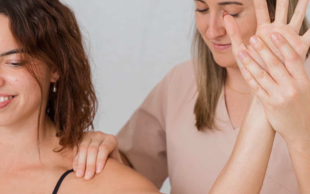
Role of Physiotherapy
Physiotherapy is fundamental to recovery. Its goals are to:
- Relieve pain and reduce inflammation (manual therapy, modalities, lymphatic drainage).
- Improve shoulder stability and range of motion.
- Restore the strength and endurance of the shoulder muscles.
- Prevent recurrence by retraining posture and movement control.

Conclusion
A glenohumeral dislocation is a treatable injury. From safe medical reduction to targeted physiotherapy, each step is essential to regaining a pain-free shoulder, restoring full mobility, and rebuilding confidence in the joint.
Recent Comments